Cardiovascular Imaging Services
Our specialists use state-of-the-art imaging to give you a clear, accurate picture of your heart's health.
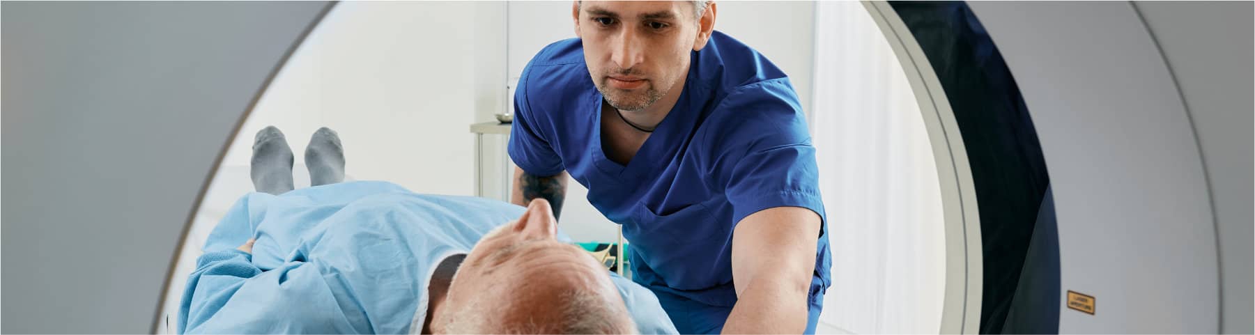
Cardiovascular imaging is essential to managing cardiac disorders safely and effectively. At Stony Brook Heart Institute, our board-certified experts deliver advanced, low-radiation diagnostics in accredited labs. We were among the first in the U.S. to offer 3D transesophageal echocardiography and continue to lead in imaging innovation.
Our team works closely with your care providers to ensure precise diagnosis and treatment planning.
Conditions Treated
Using advanced imaging, our cardiologists can see the detailed structure and function of your heart and blood vessels. These noninvasive procedures help your physician diagnose a wide range of heart and vascular conditions, including:
- Coronary Artery Disease (CAD)
- Heart valve disorders
- Congenital heart defects
- Aneurysms
- Heart failure
- Arrhythmias
- Peripheral Artery Disease (PAD)
- Cardiomyopathy
- Pulmonary hypertension
- Pericardial diseases
- Blood clots
- Aortic diseases
Cardiovascular Imaging Services
The Cardiovascular Imaging Services at Stony Brook Heart Institute offers the latest technology in a comfortable, patient-focused environment. We specialize in using the lowest possible radiation dose, which is particularly important for our younger and female patients. Our comprehensive imaging services include:
- 3D Transthoracic and Transesophageal Echocardiography
- Cardiac Computed Tomography (CT), including calcium scoring and noninvasive coronary angiography
- Cardiac Magnetic Resonance Imaging (MRI), the gold standard for evaluating heart structure, size and function
- Nuclear Imaging Studies (MUGA, PET)
- Stress testing
- Advanced techniques like Speckle Imaging, Tissue Doppler and Cardiac Resynchronization Imaging (CRT)
We also perform intracardiac and transesophageal echocardiography to guide innovative procedures in the electrophysiology lab, catheterization lab and operating room.
Our Team
Our team offers decades of experience, insight from cutting-edge research and a commitment to providing a positive patient experience to you. All our physicians are board-certified in cardiology and at least one imaging subspecialty.
Frequently Asked Questions
A 320-slice computed tomography (CT) scanner is an advanced, faster, and more precise imaging tool. While traditional CT scanners take 10 to 20 seconds to capture an image of the heart, this cutting-edge technology does it in less than one second — within a single heartbeat. Additionally, it uses an extremely low dose of radiation, thanks to the innovative techniques employed at Stony Brook.
In April 2010, Stony Brook University Medical Center became the first facility on Long Island to install a 320-slice CT scanner in its emergency room. This technology is particularly valuable for patients experiencing chest pain or suspected heart attacks, enabling quicker and more accurate diagnoses when time is critical.
Speed is crucial in medical imaging for several reasons. First, faster scans reduce the patient’s exposure to radiation, making the procedure safer. Second, a quicker scan — completed in just one second — produces more accurate images. This is because patients must remain completely still during the scan, avoiding actions like breathing, blinking, or swallowing. It’s much easier to stay still for one second compared to the 10 to 20 seconds required by less advanced machines. If a patient moves during a longer scan, the process may need to be repeated, leading to additional radiation exposure. Faster imaging minimizes these risks while improving accuracy and patient comfort.
The emergency room (ER) is an ideal location for a super-fast CT scanner because chest pain is one of the most common reasons people visit the hospital, accounting for nearly six million ER visits annually across the United States. Diagnosing heart attacks can be challenging, but the 320-slice CT scanner allows for rapid and accurate detection, enabling immediate and appropriate treatment.
Traditional ER protocols for chest pain often follow a lengthy, step-by-step process. Patients typically undergo an EKG (electrocardiogram) first. If a blockage is detected, they are sent to the cardiac catheterization lab. If the EKG is normal, a blood test is performed to check for heart damage. If the results are inconclusive, patients may need to stay in the hospital for additional blood tests six hours later. If those results remain unclear, a stress test is scheduled, often requiring an overnight stay and further delays. This process can take up to 24 hours, causing significant stress and disruption.
At Stony Brook, the 320-slice CT scanner streamlines this process. After the initial blood test, patients can undergo a CT scan. If the scan is negative, they can be discharged within hours, confident they did not have a heart attack. The scanner can also detect other conditions, such as blockages or soft plaque, providing early insights into potential heart disease. This approach reduces the total time in the ER to about six hours—more than two-thirds faster than traditional methods—while minimizing stress and disruption to patients' lives.
Stony Brook stands out due to its dedication to cutting-edge technology, its commitment to serving the community, and the expertise of the team managing the program. The medical center prioritized the integration of advanced imaging technology to improve patient care and outcomes.
Since 2009, the Heart Institute has built a comprehensive program to fully utilize CT scanner technology. This included training nurses, technologists, physician assistants, and support staff, as well as implementing specialized protocols to maximize its effectiveness. By performing cardiac CT scans daily, the team documented significant improvements in patient care, outcomes, and cost efficiency.
The decision to install a 320-slice CT scanner in the ER was driven by its ability to transform how chest pain is assessed. This technology has revolutionized emergency care at Stony Brook, offering faster, more accurate diagnoses and better outcomes for patients.
Absolutely. At Stony Brook University Medical Center, every cardiac scan is either done by me or in the presence of a physician trained by me in low-dose cardiac imaging. Since I joined Stony Brook in January 2009, I have been available 12 hours a day, 7 days a week to either perform or oversee and read every scan.
It is critically important to have an experienced physician perform and read the scan versus a technologist. It takes years of study to perfect the precise nuances of delivering the lowest dose possible for each individual patient. It is a very particular specialization that takes into account a number of factors. A trained eye and extensive experience are necessary for optimal results.